
This blog is soon to be moving and all it's contents with it.
From February 2014 the domain name will be www.davidlower.ca
Please continue to visit me there.
Happy New Year to everyone.
Shin
Splints & Achilles Tendonitis:
 |
| The muscle in the front is the tibalis anterior. |
 |
| Triceps surae is the attachment of the gastronemius & soleus muscles to the achilles tendon. The soleus is the muscle seen here. |
Achilles tendonitis is simply ‘inflammation
of the achilles tendon or triceps surae’ (pic below). For reasons to be discussed shortly
the achilles tendon becomes under increasing stress and starts to develop micro
tears. So when the muscle is engaged again and again in activities such as
running, these micro tears become more and more irritated and inflamed. This is
the pain felt at the bottom of the leg.
Here are the in depth reasons behind
achilles tendonitis and shin splints:
The
tibia bone:
The bone (pic above) is designed to transmit forces out
of the body via the foot. The outer layer of the bone, the periosteum, is
highly innervated and therefore very pain sensitive. When tension from the
muscles pulls on the bone, the periosteum develops very minute tears on the
surface and becomes irritated and inflamed. This inflammation combined with the
increased pulling action of the muscles increases the actual pressure inside
the bone. So every time you run or your foot impacts the ground, force shoot up the ankle joint into the tibia bone. Because the tibia bone is already
highly pressurised this extra force only increases the stress in the bone
further, hence the increase in pain. Repetitive actions like this, without
treatment, can often lead to muscle tears, muscle ruptures or stress fractures.
After a few years of dealing with this injury I have developed a technique to
actually reduce the tension in the bone. Within my
treatment I can also use my Bio-energy stimulation machine (B-E-St) to reduce the inflammation.
Toe
off - the last action of walking:
 |
| The toe off phase is shown by the foot on the right. |
 If the toe is restricted, it means you will
be lifting the foot off the ground at a slightly earlier stage than normal.
Consequently the tibialis anterior and calf muscles (triceps surae) have to
engage earlier to lift the foot off the ground. Your centre of gravity will not
reach the point where your body weight is directly over the standing leg. This
means you will be leaning slightly backwards and off balance as your body
weight passes behind the standing hip. The toe off phase is there to push the
body forward enough so that it sits directly on top of the standing hip, knee
and ankle. That means the forces generated by gravity will pass though all
these joints evenly and out of the body as the other leg swings forward to
complete your stride. Because the centre of gravity no longer sits directly
over the standing leg, but rather just behind it, the trunk muscles
(abdominals) and thoracic muscles have to contract to bring the upper torso
forward to give you enough balance to stand on one leg.
If the toe is restricted, it means you will
be lifting the foot off the ground at a slightly earlier stage than normal.
Consequently the tibialis anterior and calf muscles (triceps surae) have to
engage earlier to lift the foot off the ground. Your centre of gravity will not
reach the point where your body weight is directly over the standing leg. This
means you will be leaning slightly backwards and off balance as your body
weight passes behind the standing hip. The toe off phase is there to push the
body forward enough so that it sits directly on top of the standing hip, knee
and ankle. That means the forces generated by gravity will pass though all
these joints evenly and out of the body as the other leg swings forward to
complete your stride. Because the centre of gravity no longer sits directly
over the standing leg, but rather just behind it, the trunk muscles
(abdominals) and thoracic muscles have to contract to bring the upper torso
forward to give you enough balance to stand on one leg.
So what is the actual consequence of
engaging the tibialis anterior and triceps surae muscles at an earlier stage
during your run? Normally when you have a smooth ‘toe off’ phase the actual
motion of the body naturally enables the foot, knee and hip to work with less
effort because the motion of the actual stride aides in the contraction of the
various muscles. However by having to engage earlier, the muscles mentioned
have to generate this motion themselves and of course this means the muscles
generate a much greater contraction and energy output. Repeat this continually
during a run over a period of time and the stress will gradually build. This
principle is also equally applied to achilles tendonitis. The triceps surae are
working harder and the achilles tendon gradually develops micro tears due to
the high stress demand. Inflammation occurs and pain results.
The other aspect of having a restricted
‘toe’ off’ phase means your stride will also decrease. This is because the
opposite leg has had to finish the swing phase earlier to compensate for the
earlier lifting from the toe off phase. If it didn’t do this then both feet
would be in the air at the same time and this is not possible when walking.
Running is slightly different because both feet are usually off the ground, but
the principle here still remains the same. The leg will compensate. Having a
shorter stride obviously means the effectiveness of your performance is
reduced.
Bunions, blisters, corns/calluses, halux
valgus, hammer toe, arthritis, bruising, toe nail injuries (often when running
the toe nail can peel off) and verrucas will all, in their own way, create some
sort of big toe restriction.
Ankle
restrictions via the mighty talus bone:
 The talus is another part of the foot that
must be checked in any running injuries. The talus is the one bone (pic to the right) that all
forces from the body travel through to eventually disperse throughout the rest
of the foot and then out into the ground. The tibia and fibula bone connect
directly to the talus giving the ankle joint 2 predominant movements – plantar
flexion and dorsiflexion (pic below). To simplify, this is the swinging motion of the foot.
To achieve this effectively the talus rotates forwards and backwards within the
tibia and fibula joint complex. However, it is possible for the talus to be
shunted forward (anterior) or backwards (posterior) (pics below). If the talus gets stuck
anteriorly then the movement of dorsiflexion (pic below) becomes somewhat reduced and if it
is stuck posteriorly then plantar flexion becomes reduced. This consequently
reduces the overall movement of the ankle joint in either direction and will
result in very similar patterns as described above in toe off. The stride will
reduce, the centre of gravity becomes displaced and the muscles work harder to
lift the foot off the ground.
The talus is another part of the foot that
must be checked in any running injuries. The talus is the one bone (pic to the right) that all
forces from the body travel through to eventually disperse throughout the rest
of the foot and then out into the ground. The tibia and fibula bone connect
directly to the talus giving the ankle joint 2 predominant movements – plantar
flexion and dorsiflexion (pic below). To simplify, this is the swinging motion of the foot.
To achieve this effectively the talus rotates forwards and backwards within the
tibia and fibula joint complex. However, it is possible for the talus to be
shunted forward (anterior) or backwards (posterior) (pics below). If the talus gets stuck
anteriorly then the movement of dorsiflexion (pic below) becomes somewhat reduced and if it
is stuck posteriorly then plantar flexion becomes reduced. This consequently
reduces the overall movement of the ankle joint in either direction and will
result in very similar patterns as described above in toe off. The stride will
reduce, the centre of gravity becomes displaced and the muscles work harder to
lift the foot off the ground. |
| Dorsi flexion is when the toes point up. |
The other aspect of talus restrictions is the distribution of force. The talus is designed to sit in the middle of the tibia/fibula complex. Therefore it is important that the force of gravity travelling from the body and down the leg travels directly through the talus. Whether the talus is restricted
anteriorly or posteriorly only means the force will disperse elsewhere in the foot, often in a non-physiological way.

An anterior restriction will result in the force travelling slightly behind the talus (green line in the pic on the right) and out through the calcaneus. (The red cross represents where the force will end). The natural motion of the talus is to slide forward on top of the calcaneus and into the connecting navicular and cuneiforms bones (purple and brown bones). With the medial arch linking all these bones together via the plantar fascia, the whole process acts like a suspension system. The navicular and cuneiforms act like a breaking system as everything compresses and bunches together. The medial arch behaves like a dampening spring to absorb nearly all the shock. However if the force passes behind the talus and straight into the calcaneus then this whole suspension system is missed and so the effect of this is to have a harder impact on the ground, which translates into greater force travelling back up into the tibia. Repeat this over time and it is no wonder the tibia bone becomes stressed. Remember also, when the foot hits the ground the achilles surae and tibialis posterior muscles are already in contraction to maintain plantar flexion as the heel hits the ground. Instantly on impact the tibialis anterior muscle contracts to decelerate the motion of the fore foot as it hits the ground. If the force or shock travelling up the bone is harder than normal, the contracted muscles will be put under greater tension because they have to work harder.
 Consequently if the talus is restricted
posteriorly (pic just below) then the force of gravity passes in front of the talus and straight
down into the medial arch mechanism, missing the forward motion of the talus on
the calcaneus and the resulting bunching together of the navicular and
cuneiforms. In time this stresses the medial arch, resulting in many possible
foot injuries, such as plantar fasciitis. However the tibialis posterior and
tibialis anterior muscles both have strong attachments into the under-surface
of the foot, into the medial arch itself. Stress the medial arch and these
muscles become naturally elongated and start to behave like contracted muscles.
In time they will become weaker as they acclimatise to the elongation stress.
When these two muscles become stressed they tighten and create an overall
increased pull on the tibia bone – shin splints. This then leads us nicely onto
what is commonly talked about – overpronation.
Consequently if the talus is restricted
posteriorly (pic just below) then the force of gravity passes in front of the talus and straight
down into the medial arch mechanism, missing the forward motion of the talus on
the calcaneus and the resulting bunching together of the navicular and
cuneiforms. In time this stresses the medial arch, resulting in many possible
foot injuries, such as plantar fasciitis. However the tibialis posterior and
tibialis anterior muscles both have strong attachments into the under-surface
of the foot, into the medial arch itself. Stress the medial arch and these
muscles become naturally elongated and start to behave like contracted muscles.
In time they will become weaker as they acclimatise to the elongation stress.
When these two muscles become stressed they tighten and create an overall
increased pull on the tibia bone – shin splints. This then leads us nicely onto
what is commonly talked about – overpronation.
What
is over-pronation and supination?

 |
| Medial arch. Looking at the foot from the inside. |
There are 3 places this force tends to go.
The heel (calcaneus), the big toe (metatarsophalangeal joint MTP1) or the
transverse arch.
The calcaneus bone has no shock absorption
except the fat pad underneath it. Repeated stress in this area will likely lead
to plantar fasciitis or heal spur type injuries.
During the normal walking cycle the heel
hits the floor first, following to the outside of the foot where it reaches the
little toe (this is called the lateral roll). From here the natural motion and
weight travels across the transverse arch to the big toe (this is called the
medial roll) and then lastly to toe off. When the medial arch has collapsed the
lateral role of the foot during this walking cycle will be either partially or
completely missed. So instead of having a lateral role movement the force just
travels straight across the foot to the MTP1 joint, hitting it more directly
and more forcefully. This will lead to chronic stiffness of the big toe and reduced
‘toe off’ function, discussed above. Additionally when the arch has collapsed
you can develop an unnatural inward rotation or twisting of the MTP1 joint,
albeit it a mild one and this leads to the inner part of the joint taking the
biggest brunt of the force. Repeated stress and offloading of force here can
lead to painful calluses, bunions and halux valgus type injuries.
 |
| Longitudinal arch = medial arch. |
Future installment:
I am currently in the process of writing about knee mechanics and their impact on these injuries. I will also look at the fascial chains running through the leg and how the viscera play their role. I hope to get this released soon. Thanks.

Wear the correct shoes:
When looking for the correct shoes it can be quite a
jungle out there with so many different types to choose from. Shortly I’ll give
a list of the top shoe brands out there. Visiting their individual website will
give you a lot of information about the best shoe for you. As a general rule
there are categories for the type of terrain you are using; such as trail,
mountain, road, race and long distance running. Within these categories you
also have the type of fit you need depending on the severity of pronation or
supination you have, plus other special requirements you may have. Lastly there
is the type of shoe to suit your running style whether it be heel strike,
midfoot strike or forefoot strike.

All the different shoe companies have many patented
technologies that all sound highly intelligent and impressive, but whatever it
is they say, you want to make sure you check and consider the following with
regards to your own feet.
Dorsiflexion:

Check the amount of dorsiflexion you can achieve and how stiff this movement feels to you. You should be able to achieve 10 degrees of dorsiflexion in a non-weight bearing seated position (you may need someone to help you). Also note how stiff it feels to push your foot into full dorsiflexion.
Determining this will help you understand how high a heel
you want. Personally I think modern running shoes are developing far too high a
heel and this can lead to something known as ankle equinus. Ankle equinus is
the medical term given to shortening of the achilles tendon leading to
reduction of dorsiflexion in the foot. So running with a high heel can lead to
a shortening of the achilles tendon. Long term shortening of a tendon like this
will undoubtedly lead to achilles tendonitis and/or shin splints. This is what
happened to me. I started wearing asics kayano and within a few weeks I
developed very painful shin splints in both legs. I believe this particular model of shoe had too high a heel. HOWEVER if you have a reduced
dorsiflexion movement or a stiff movement in general, it may help to start off with higher
than normal heels and gradually acclimatise over time to a reduced heel size.
Having the slight heel increase will help with supporting your reduced
dorsiflexion and prevent overstrain to the achilles tendon.
Determining the degree of heel raise you want to look for
phrases such as, heel to toe differential, heel to toe offset or heel to toe
lift etc.
Ankle Support:

 If you have sustained any form of ankle sprain in your life
you should consider this when buying your shoe. Lateral sprains (outside of the
ankle) are the most common with medial (inside of the ankle) being the most
severe. The subtalar joint (pic directly right, formed by the talus in grey and the calcaneous below), which I haven’t mentioned up until this point,
produces the small movements called inversion and eversion. When ligaments are
torn, which is what a sprain is, they are permanently stretched. Ligaments do
not contain any elastic properties, so any unnatural force that causes the
ligament to elongate will mean that ligament is permanently elongated because
there is no elasticity to bring the ligament back to its original position.
This means the ligament no longer functions properly to support the subtalar
joint and therefore the inversion/eversion movements can increase.
If you have sustained any form of ankle sprain in your life
you should consider this when buying your shoe. Lateral sprains (outside of the
ankle) are the most common with medial (inside of the ankle) being the most
severe. The subtalar joint (pic directly right, formed by the talus in grey and the calcaneous below), which I haven’t mentioned up until this point,
produces the small movements called inversion and eversion. When ligaments are
torn, which is what a sprain is, they are permanently stretched. Ligaments do
not contain any elastic properties, so any unnatural force that causes the
ligament to elongate will mean that ligament is permanently elongated because
there is no elasticity to bring the ligament back to its original position.
This means the ligament no longer functions properly to support the subtalar
joint and therefore the inversion/eversion movements can increase.
So it is important to consider solid ankle support to reduce
this increased inversion/eversion movement. If this movement is severe in some
people they are more prone to spraining their ankle again when running. Additionally
your leg muscles will have to work harder to prevent and stop the ankle
producing too much of this movement and maintain balance.
Over pronation:
 |
| A. Normal foot, B. Pronation (collapsed arch or flat foot), C. Supination (high arch). |
With a wet foot, walk on an A4 piece of paper (preferably a
coloured piece) and see what happens. You foot should look something like A on the picture to the right. If you foot looks like B then you are over pronated and if your foot looks like C then you have high arches. Most people have normal to
over pronated arches. This means you will want to look for a shoe to support
your medial arch to the degree to which it has collapsed. High arches on the
other hand mean you will want support for the toes, especially at the little
toe area. This is where most of the pressure will go as the spring mechanism of
the medial arch is bypassed. So you want shoes that accommodate this. This is often
called mid sole cushioning or lateral arch cushioning.
Other considerations:
Shoe companies often talk about foot spread or how wide the
shoe is. This is also beneficial to consider to you that have quite wide or
thin feet. When you run your feet will naturally expand and so you will want
shoes that can accommodate this expansion, rather than squashing your toes
together during your run. With this you can also consider any calluses or
bunions you may have too. You want make sure your shoes will have a good mid
sole width (that is often where the shoes laces begin at the bottom of the shoe).
It is also worth noting that not all two feet are actually
the same. I for example have quite a reduced arch on my right foot as opposed
to a normal arch on my left foot.
Here are some of the top running shoe brands and like I say
their websites are often worth checking out for extra information.
Asics
Newton Gravity
Salomon
New Balance
Brooks
Saucony
Hoka one one
Vibram 5 fingers
Nike
Addidas
For months now I
have been writing this article about scar tissue. I have found quite a lot of information, but
as usual nothing on a deeper level. A lot of books and websites will say scar
tissue can create blockages, but I want to know why and how. So I hope what I
have written will provide some of these answers.
What is scar tissue?
In my line of
work we tend to use other phrases like abdominal adhesions or fibrosis to
accurately explain scar tissue.
Abdominal adhesions are pretty specific to internal workings of the
body, usually linking an organ to an organ or tissue to an organ. Adhesions are
fibrous bands that connect one tissue to another tissue, forming a connection
that physiological shouldn’t be there. These bands are fibrous, meaning they
are tougher and less flexible than their surrounding tissues to which they make
the connection. Hence the phrase ‘fibrosis’.
When adhesions
form between two different organs it can create a tethering, much like an
anchor on a boat. One organ becomes partially fixed and therefore permanently
struggles to expanded, stretch, move or glide. Sometimes a tether can also
behave like a torsion and actually constrict an organ, creating a physical
blockage. This is especially true in tubular organs. Within both of these
examples, movement and physiological function is lost. I want to focus on the
movement aspect first. Reduced movement in anything, especially with regard to
the body, is a bad thing. Whenever there is a reduction in movement there is
also a reduction in nutrient supply and reduction to toxin expulsion. Too much
toxin build or not enough nutrient supply can lead to cell death. Any form of
death in the body automatically triggers an inflammatory response. Similarly if
stagnicity is present then there is a much greater chance of infection.
Stagnicity indicates a lack of sufficient blood supply and therefore a lack of
immune response to the targeted area via the blood supply.
So what
happens?
In the case of surgery, the natural healing
process is kick started by inflammation. Inflammation brings all the materials
needed for repair and clears the site from harmful microbes. Fibrin is then laid
down, acting like glue and eventually creates a fibrosis. Collagen is the last
substance to be introduced.
 Collagen is
present in pretty much every tissue of the body and is usually laid down in a
random, chaotic pattern [picture to the left].
Collagen is
present in pretty much every tissue of the body and is usually laid down in a
random, chaotic pattern [picture to the left].
However in scar tissue, collagen is laid down in
heavy concentrations in a linear pattern or a straight line pattern. This is
what gives scar tissue that white smooth linear look [pictures below].

When collagen is
laid down in thick fibrous bundles the blood supply becomes very insufficient. Collagen
is very inflexible and together with the poor blood supply, it makes an area of
scar tissue quite lifeless. So the vitality that was once present in the
healthy tissue pre trauma (operation) becomes a rigid, tense and almost
lifeless area.
If you think that
collagen is the primary ingredient in bones and ligaments you will begin to
understand the far reaching consequences of putting a material like this
directly into a healthy functioning tissue, especially an organ.
 Here is an
example to try and explain the consequences of abdominal adhesions:
Here is an
example to try and explain the consequences of abdominal adhesions:Imagine taking a normal balloon, it blows up normally and evenly. Now imagine putting a strip of duck tape on it, like a plaster. When you blow up the balloon again, everywhere expands except the area of duck tape, which stays fixed. The balloon then expands around the duck tape and forms an asymmetrical blob. It no longer looks like a normal balloon. This is how scar tissue or collagen functions when laid down in places not originally designed for that purpose. This leads on to the model of Osteopathy I’ve tried to explain before. Everything is motion, moving, fluid, and breathing. Put a fixed, fairly lifeless structure into this system where it is not designed to be and it will start to become a stress on the system, all bit a minor stress to begin with.
So
what are the consequences of surgery?
When surgery is performed external air will
get into the abdomen and this dries out the natural lubrication and viscosity
of the various abdominal organs and tissues. Often blood is split and becomes
sticky, producing the same consequences. When the layers become dry or lack
lubrication, this will increase the friction of movement between the layers. The
various layers are now pinned together, and the once free movement upon each
other, is now dictated by the scar tissue. They can no longer slide and glide
upon each other. This increases the friction between the various layers.
If you take two glass panels and put water between their two surfaces and slide them over each over, it is effortless. Remove the water and slide the glass again and it becomes a lot less smooth. This is the principle here. The viscosity changes after trauma or surgery and so in time this can add small repetitive stresses. [see video below - laparoscopic appendectomy].
If you take two glass panels and put water between their two surfaces and slide them over each over, it is effortless. Remove the water and slide the glass again and it becomes a lot less smooth. This is the principle here. The viscosity changes after trauma or surgery and so in time this can add small repetitive stresses. [see video below - laparoscopic appendectomy].
I want to thank 'Kiplinght' for permission to use his video.
The other aspect here to consider is when an organ becomes unnaturally fixated its axis of movement changes. This has far reaching effects on the other organs because the organs all rotate and move to a certain rhythm. The ascending colon has attachments to the kidney, duodenum and liver. All these organs move and they move together, effortlessly and cohesively. When the axis of movement changes, say at the ascending colon for example, then instantly the colon will pull away from its natural pattern of movement and therefore pull on the other 3 organs just mentioned. In time this can put stress on these organs and they can go into dysfunction too.
If the axis of movement to which an organ
rotates around changes, then this can also alter the dynamics of the mechanoreceptors.
Mechanoreceptors are neurons that pick up changes to pressure or stretch. So if
a stretch or distortion exists that shouldn’t exist, then this could
unnaturally trigger the mechanoreceptors. The feedback goes to the spine/brain and
comes back resulting in spasms to the muscles of the organ.
In the stomach, when the mechanoreceptors
are triggered, the stomach begins to release hydrochloric acid (HCL). So if
there is a fixation on the stomach, due to some external input, the mechanoreceptors
can fire and produce more HCL acid. This increases the acid production which in
time can lead to possible ulcerations or a hiatus hernia. The mechanoreceptors
are designed in this case to be triggered when the stomach is becoming
stretched as a result of food entering it, not through other stimuli.
So here comes the million dollar question; can scar
tissue be treated?
Yes I believe scar tissue can be treated
after surgery. But first we must look at scar tissue from a slightly broader perspective.
When the body heals from a wound, it usually has some consequences that stick
around after the healing processes have finished. The wound site is often
bigger than the resultant collagen scar tissue. There is nearly always an
affected area around the scar that usually presents with stiffness, reduced
vitality and reduced function. It is this area that is treatable.
By pulling, stretching, twisting or lifting
an organ we can release the tensions spreading in and around the site of scar
tissue. During an appendectomy a cut is made down to the large intestine from
the skin, through a fat layer, through fascial layers, through the abdominal muscles
and then finally into the abdominal cavity (recently they have started using
laparoscopy, which minimalises scarring). The cut to reach the Cecum is
actually relatively small and often the resulting scar tissue is small. However
we often find that the entire Cecum and ascending colon is fixated and has lost
its functional movement. So the area of injury or trauma is no longer actually
focused towards to the site of scar tissue. The aim would therefore to be to
lift the Cecum off the posterior abdominal wall, stretch the iliocecal
ligaments located inferiorly and to also stretch the ascending colon. This
often nearly always clears up the problem and functional movement returns to
almost normal. I do not believe however that the physical scar tissue, made of
collagen is treatable. But this is just my opinion.

Bag It!!!
I recently watched a very profound documentary on plastic called 'Bag It'. It talks about how we are struggling to deal with the waste of plastic, the problems we are facing with global consumerism and the health effects of using plastic in our foods. Although its delivery and presentation was ok, the underlying story and information was fantastic. I would strongly advise purchasing this DVD, albeit an expensive one, and give your support to the works of this organisation. As usual the link is above.
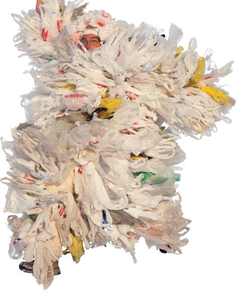

The North Pacific Gyre
I have recently been reading about some issues regarding plastic and general waste. During my search I found the above expression, the north pacific gyre. I was shocked at what I discovered.
This is a huge area in the north pacific ocean that has accumulated a huge volume of trash, mostly plastic as it does not biodegrade. Check out the link above for more information, it's scary stuff.

Principles of scar tissue:
Firstly I want to highlight a principle I think is the most important in my line of work. If you read ‘what is Osteopathy’ in the ‘Osteopathy’ tab on this blog you will see I have written about what I believe Osteopathy is. There I remark on the principle of motion and that the body is constantly moving. It is very important to have this in the back of your mind as I try to explain the consequences of an appendectomy or scar tissue in general.
When the appendix is removed an incision is made in the right lower quadrant of the abdomen. This cut slices through a lot of different layers of tissue. The appendix is then cut away and the large intestine is stitched up. More recently they have started using a laparoscopic procedure to remove the appendix – link here.
The point I want to highlight though, is when the various incisions are stitched up scar tissue will form. Scar tissue is non flexible, it doesn’t stretch. All the other layers and tissues that have been cut through can stretch; they expand and relax with the various motions of the body. They adapt, they live and by this I mean there is cellular exchange and interaction. Scar tissue however cannot move or stretch, it is considered ‘dead tissue’. I’ll explain more about scar tissue in a different blog.
When you have something that is not flexible blend into something that is flexible it will create tension throughout the tissue that is trying to expand, it will act like an anchor. When this occurs constantly every minute of every day, stress will eventually build in the surrounding tissues in connection with the scar tissue. A good picture to paint here is normal tissue acts like a healthy runner. It can jog for hours not draining the system. When you get scar tissue the healthy tissue continues to jog, but now it is dragging a car tire behind it. Stress builds, compensation occurs, function is reduced and ultimately injury can occur.
Another feature of scar tissue is the production of adhesions. In very simple terms this basically means that different layers of healthy tissue can stick together and this stops the natural function and mobility of that tissue.
On an interesting note, injuries can develop years later after surgery or any other kind of trauma. Just because there are no symptoms now, it does not mean compensation and adaptation patterns are not already occurring due to the increased task load of that tissue. It is also common to have an apparently non-symptomatic scar tissue trigger into a symptomatic problem when another injury/trauma in the body occurs later on. This is why someone can often develop a shoulder problem years later after a pelvis or leg injury for example. Years of compensation build over time and then a new trigger in the form of a new injury/trauma can occur.
Conversely, do not fear that all of a sudden you are going to get serious problems just because you may have had surgery in the past. I am merely highlighting why sometimes things can suddenly become symptomatic years down the line, when apparently there has been no problem.
So what can happen after an appendectomy?
 With an appendectomy, the cecum (beginning of the large intestine) can become fixed to the back wall of the abdomen and the back wall, in this case, would be the transverses abdominis or iliacus muscle. The cecum is also the junction between the small and large intestine and in some woman it attaches to the right ovary via the ligament of Cleyet. When I talk about attachments I'm referring to either ligament, fascial or membrane connects between various organs or bodily structures.
Higher up as part of the ascending colon it attaches to the duodenum (the beginning of the small intestines), the right kidney, liver, gallbladder and finishing up fixing to the diaphragm. I highlight all these attachments because these can become potential areas of compensation, irritation or sites of 'injury'/symptoms. So when the inherent movement of the cecum has stopped due to fixation caused by the scar tissue it creates what I call drag. This is another principle resulting from scar tissue. This drag almost acts like a gravity field. Not only is the inflexibility of the scar tissue causing tension to spread through the structure, it also causes other nearby structures associated with it to get pulled towards the scar tissue or fixation.
[The picture above shows all the attachments of the large colon - indicated by arrows].
Knee and ankle problems can develop:
 The cecum attaches to the iliacus muscle of the pelvis, which works to produce flexion and internal rotation of the hip. If the cecum becomes stuck (it's lost its inherent movement) it can cause the iliacus muscle to pull towards the area of fixation. It is almost like the two structures start to act as a single structure. Ultimately this causes the muscle to become chronically contracted and the hip to become more and more internally rotated and flexed. This will lead to internal rotation of the femur and then internal rotation of the knee joint. The knee joint will also stay further in flexion. Both these actions result in the tibia shifting further forward on the ankle joint and inwards towards the medial arch of the foot. This can predispose to stress on the medial arch, which over time can lead to collapsing of that arch known as over pronation. Conditions like plantar fascitis and heel spurs can develop. So with just this simple chain we can see the potential of knee, ankle and foot problems occurring just from a fixation due to an appendectomy. Remember these are all potentials and NOT definite compensations that will happen to everyone that has had an appendectomy.
Neck problems can develop:
 So going back to the other structures that attach directly or indirectly to the cecum. I'm going to leave out the duodenal and kidney attachments here otherwise I'll end up writing a book rather than a blog. But let's focus on the liver and diaphragm attachments. So through fixation of the cecum, the ascending colon is gradually being drawn down towards the cecum area via the drag and this directly pulls on the liver and diaphragm. Remember the image of the car tire. Suddenly the liver is having to move with an unnatural force pulling it constantly downwards towards the hip. The liver has massive attachments to the diaphragm and then with the direct attachments of the ascending colon as well we suddenly find the diaphragm is struggling too.
[The picture directly above and left shows how high the ascending colon goes, sitting directly under the liver and diaphragm. The picture to the right shows the pleura of the lungs, the grey membrane sitting on top of the red muscle (diaphragm)].
Now on the upper surface of the diaphragm you have the pleura of the lungs (the membrane that covers the lungs) which blends directly into the diaphragm. On the upper surface of the lungs, the pleura attaches to the vertebral bones of the neck via a fascia called Sibson's fascia. So with the pull of the diaphragm down, you also get a pull of the pleura, which pulls on the neck. Neck pain, stiffness and tension can occur.
I think it is important to clarify here that although I’m talking about one structure pulling on another structure and then that other structure pulling still further on another structure; it is slightly different within the body. We have to remember the body is living and everything is connected, even if it is not physical, it is still connected by blood, nerves, emotions, hormones and gravity. It is very difficult to highlight how this represents in the body via words. It is something I have just learnt to feel – perhaps it is intuition. In the medical world when we learn anatomy we cut everything down and separate everything to its basic level, so we can learn. But to grasp how the body truly works we need to build up that anatomy again and see a whole living body once more. It is very easy to think “how is it possible for the Cecum, which is all the way down near the hip to affect the neck – the two structures are so far away?” In the body these structure are not far away. In fact they are touching, but perhaps not in a direct way, but rather in an indirect way.
Low back pain is common:
Here I am going to talk about a structure called ‘toldt’s fascia’. This structure is a membrane type structure or for understandings sake let’s just say it acts like a piece of ‘cling film’ or ‘shrink wrap’. This membrane starts from the spine, spreads out and wraps around the ascending colon to then travel back to the spine again. So when the colon becomes fixed, this can lead to stress and tightness spreading through the fascia, resulting in a pull on the spine. Over a prolonged period of time it puts a greater demand on the spine and it gradually stiffens up. This is what happened to the patient I had recently; refer to the ‘Bob the builder story’ on my blog. The Cecum became fixated due to the scar tissue and adhesions. This created tension to spread through toldt’s fascia and then lead to tightness and pain in the lower back. This is why after 4 treatments of manipulation, stretching and pulling on the spine, no improvement was achieved; because all the while his colon was still fixed causing the tightness in the spine to remain.
Signs and symptoms of cecum fixation:
The large intestine works to absorb the remaining water (about 10%) and take in the remaining vitamins (mainly E, K & B12). Interestingly the large colon has a very high percentage of bacteria which ferments the food (this is very healthy for the body). This increases gases and therefore smell, but this is normal.
Depending on whether the fixation of the cecum is in a state of irritation or a state of passivity will depend of the symptom. An irritation is more an excitatory response and the speed of which peristalsis occurs increases. Therefore diaherria and dehydration can occur, simply because the stools pass through too quickly for the water to be absorbed.
If it is passive then think stagnicity. Constipation, bloating and flatulence can occur because the stools are not passing through quick enough, all the water is absorbed leading to harder, firmer stools, which are difficult to pass. Stagnicity leads to longer fermentation periods and this obviously produces more gas and bloating, also known as trapped wind, which actually is extremely painful.

Bob the builder:
I treated a recent patient of mine, Bob (not his real name of course), for a very typical low back problem. After four treatments of the normal stretching, pulling, manipulating, pushing and bending he improved to perhaps 60-70%. We unfortunately could not breach that final 30%, which would have returned him back to his normal quality of life. He has tried the usual therapies in the past and they too were getting the same sort of results. We were all hitting an invisible wall it seemed.
So on one of his treatment I sat down pondering his situation/condition. He was your typical stiff builder type with very little flexibility and had been suffering with low back pain on and off for 1-2 years. No neurological symptoms or anything else untoward. I had been treating a lot in his upper back, rib cage and breathing mechanics too. On the third treatment I looked at his legs and feet. Always there was an improvement, but never beyond that 70% mark.
 On the fifth treatment I was working on his lumbar spine (low back) when I noticed how stiff it was considering he had had four treatments earlier of stretching, bending and all the above. It suddenly struck me how odd this was and so I decided to re-read his medical notes, starting with the list of his operations and medical conditions. This was when I came across his appendectomy (removal of his appendix) some years earlier. So I sat him down and checked his abdomen. Sure enough he had a lot of tension around the cecum (beginning of large intestine and appendix. I treated this area very simply and then stretched toldt’s fascia on both sides. When I had finished completing these 2 easy techniques the patient remarked on how relaxed his back felt.
 [The picture on the left is an overview of the large and small intestines. The picture below is a close up of the cecum and appendix; the appendix being held by the metal tweezers].
Sure enough he came back to his next treatment nearly 100% better; the best he had felt since the beginning of his low back problems. Although this has been a great success, it has also been a solid reflection. I almost missed this because I assumed he just needed a good stretch and manipulation, which in most cases is exactly what this type of patient needs. But it was that feeling I felt in his back that just made me very uncomfortable, as if something was wrong. A back that is stretched and manipulated regularly for a few weeks should change in vitality and composition. This back however stayed the same since the first treatment. Something completely different needed to be done.

In another blog that will follow shortly in the next few days I will explain the mechanics of what happens to the body after an appendectomy and the reasoning to why this caused Bob a lot of back pain for a year.

Here is a useful tip I learnt the other day. Take a spoonful of peanut butter perhaps 30-40 minutes before you do a weight training session and you will have more energy to burn. Obviously if you have allergies to peanuts then don't follow this advice.
Do you remember doing that experiement at school where you set to flame a poor innocent peanut and timed for how long the flame kept alight? Afterwards you did some maths to determine how much energy was contained within that peanut. I always remember the peanut burning for quite a long time.
 For the third time today I am completely re-writing this blog piece. I have spent all day researching the microwave and now, late in the evening, I have finally come across an article I am satisfied with. Check out the link here.
When you research and study something for so long you pick up the trends, stories, myths, arguments, research and just general know how. What I liked about this article is that the author covered all the major trends, theories and arguments and then carefully discredited them each in turn. I thought it was brilliantly written and provides an alternative to the huge number of ‘anti microwave’ reports and websites out there.
Having read so much today and picked through the various research papers I can safely say she covered the majored topics. I am actually quite relieved because this piece was turning out to be a monster to write. The other website that had a good lengthy report on the microwave, and that is anti microwave, is here. Pick through the evidence and decide for yourself. I have heard quite a few times within the health industry that the microwave is bad and well here are the facts for you to explore yourself.
 Continuing on from this theme of positive thinking, I spent a few hours researching this field and trying to find out what medical research has to say about it. To cut a long story short I found the name of, probably, the leading expert in this area. His name is Martin. E. Seligman. I personally have never heard of him up until now, but I found a website with all of his various papers and it looks very extensive. Click here:
I have not had time since to explore all of his papers, but I did buy his book which has had many good reviews. It is the picture above and the book is called; ‘Learned Optimism’. I am going to read this book and once finished, review it.
One thought I have had recently and have had many times, especially when it comes to the argument of eating healthy, is the importance of combining positive thinking with eating well. I believe nutritionists do not give this area of combining positive thinking and eating healthy enough credit or attention. This is not an attack against nutritionists, far from it. A lot of medical practitioners leave the mental state of health on the shelf sometimes, for which I too find myself guilty of sometimes.
I am convinced that there is at least a 50/50 balance between the effects of eating healthy and the effects of being positive about what you eat and the possible failings along the way. I do not have any proof for this and it is just my opinion, but for example, if I ate a healthy salad for lunch and resisted all the urges to have a cake or eat chocolate, then I am naturally going to be pleased with myself. This pleasure or satisfaction produces endorphins in our brain that numbs pain and reduces stress. Of course eating the salad and limiting the excess sugar or processed foods will create and provide the body with a fuller nutritional supply. Therefore the combined effort of eating health, but also thinking positively is optimal. This piece on endorphins was very interesting:
Further to this is the consequence of negative thinking, which I believe is extremely powerful. Say I eat that same salad, but I instead decide to eat the cake afterwards for whatever reason. The guilt and sense of failure is really strong, especially if you are self conscious about your body and believe eating healthy is the answer to getting a better image. So you have failed on this particular meal time, but the consequence of it lasts way beyond that actual meal. I believe so strongly that guilt and self-loathing is detrimental to a person’s inner well being. The internal stress that guilt and condemnation produces, I think, is on a similar level to a physical trauma. People don’t associate these feelings as an internal stress. The word stress is often over used and it is unfortunate because there are actually many different types of stress and negative thinking is amongst the strongest stresses you can put your body through. Again this is just my belief.
If is so important to learn to indentify feelings or even thoughts of failure, worthlessness, insecurity, stupidness, weakness and guilt. There are many more here I haven’t mentioned, like anxiety or anger, but I want to just concentrate on the thoughts of self. Once an ability to highlight them is created then the battle to change can really begin. It is the process of recognizing these thoughts/emotions and not letting them take root, but rather turn 180 degrees around and say the opposite to what you are thinking/feeling. One day you fail at something or in the past your parents/teachers have told you will never make it, that you will always fail. It is taking that thought, however many times in arises during the day, and then saying;
“I am not a failure, I can do this”.
“I am not a waste of space, I am somebody important”.
When we can take these steps; change to the positive is inevitable and I am convinced anybody can achieve these goals in whatever their struggle. It is important to remember at this point that we will fail at times to keep remembering the steps or may find it extremely exhausting to keep reminding ourselves to work on this process, but it is utterly essential to not beat yourself up and allow guilt to destroy the progress of your work. Stumbling and getting it wrong is always a part of learning a new thing, like walking as a baby. Just don’t be hard on yourself, give yourself a little grace. It is also vitally important to remember that, sometimes, when you have turned 180 degrees around and declared the positive, nothing seems to have changed. No new emotions can be perceived. This at times can be very de-motivating, but it is part of the process. So just keep going.
Positive thinking in this way reduces stress in our lives and it is well known through research that optimistic, ‘happy’ people are healthier and achieve more in life. They also recover better from injuries and health problems – which makes my job easier.
You can do it.


 Posts
Posts


